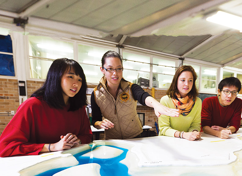
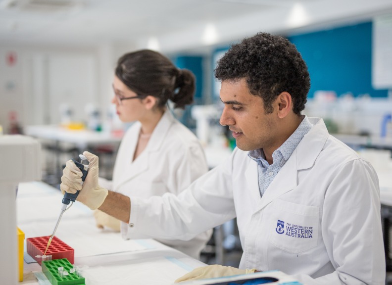
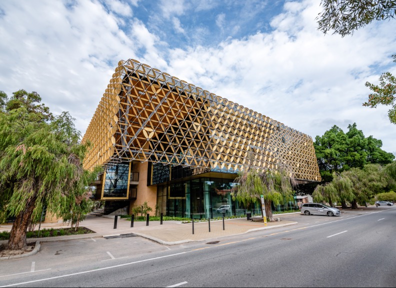
BRITElab
State-of-the-art laboratory for bioimaging and biomedical engineering research
Researchers at BRITElab (Bioimaging Research and Innovation for Translational Engineering laboratory) develop and translate novel medical imaging techniques for a range of clinical and biological applications.
The primary focus of BRITElab is the development of optical coherence elastography (OCE) techniques. OCE is an emerging imaging modality that maps depth-resolved mechanical properties of tissues at high resolution.
To perform OCE, a mechanical load is applied to the tissue and the resulting deformation is measured using optical coherence tomography. The measured deformation can be used to:
- Provide qualitative mechanical contrast of tissue by generating images of strain
- Quantify mechanical properties (eg. elasticity) by solving the forward mechanical problem
BRITElab has developed a range of OCE techniques suitable for biological applications including oncology, cell mechanics and tissue engineering. In addition the lab collaborates closely with OncoRes Medical, a UWA based start-up company, to translate these techniques from a laboratory-based method to the clinic
One of the lab’s notable achievements was developing a medical imaging technique allowing surgeons to easily locate tumour during surgery.
Founded in 2016 by Dr Brendan Kennedy, BRITElab research includes developing wearable biomedical optics devices, intraoperative surgical techniques, optical elastography, and tissue and cell mechanics.
Research publications from BRITElab can be found here
![]()
BRITELab Team Members
- Brendan Kennedy
- James Anstie
- Qi Fang
- Ranate Zilkens
- Erin Lloyd
Lab Head
Post-Docs
PhD Students
- Helen De Jong
- Ken Foo
- Matt Hepburn
- Rowan Sanderson
- Wes Allen
- Anshul Goplani
- Daniel Firth
- Johnny Choi
- Mark Mazzoni
- Wayne Adams
- William Wong
Undergraduate and Masters Students
Cross-disciplinary Collaborations
- Prof Christobel Saunders, Dr Bruce Latham and Dr Benjamin Dessauvagie
-
Approximately 1-in-4 women undergoing breast-conserving surgery require a second or third surgery due to tumour found at the edge (margin) of the excised tissue, which indicates that there might be residual tumour in the patient and is associated with a higher risk of cancer recurring.
BRITElab are collaborating with breast cancer surgeon, Prof. Christobel Saunders, and pathologists, Dr. Bruce Latham and Dr. Benjamin Dessauvagie to develop optical coherence elastography techniques to enable intraoperative assessment for tumour during breast-conserving surgery. The team are developing both bench-top systems for assessing the margin of the excised tissue and hand-held probes for detecting residual tumour in the breast cavity. The technical and clinical feasibility of the techniques are being investigated at Fiona Stanley Hospital, Western Australia.
See more research publications:
- OncoRes Medical
-
BRITElab is collaborating with OncoRes Medical in the development of intraoperative imaging technology to provide surgeons with real-time assessment of tissue microstructure. Physicians have relied on the sense of touch, manual palpation, for centuries to diagnose disease by feeling for changes in mechanical properties. One area where surgeons continue to use manual palpation is for guidance during breast cancer surgery. However, manual palpation suffers from poor resolution and is subjective, and additional surgeries are common due to the cancer not being fully removed. By providing surgeons with high-resolution images of tissue stiffness using optical coherence elastography, we aim to translate the surgeon’s sense of touch to the micro-scale, resulting in improved outcomes for patients.
Find out more about OncoRes here
- Dr Yu Suk Choi
-
Mechanical forces serve as critical regulators of healthy cellular function and tissue development. The forces and resulting deformation that a cell experiences are implicitly linked to the mechanical properties of both the cell and the surrounding extracellular environment. Cells actively sense and exert forces in their environment and convert them into a chemical response in a process known as mechanotransduction. Diseases associated with altered mechanotransduction include muscular dystrophy, cardiac myopathies, atherosclerosis and cancer.
BRITElab are collaborating with Dr Yu Suk Choi from the Stem Cell Mechanobiology Lab in the School of Human Sciences at the University of Western Australia to develop techniques to study the impact of mechanical properties on human stem cell interactions in 3D using optical coherence elastography.
See Dr Choi's research publications here
- Prof Assad Oberai
-
Collaboration with Professor Assad Oberai from the University of Southern California aims to use solutions to the inverse problem of elasticity using adjoint elasticity equations to achieve cellular resolution optical coherence micro-elastography.
Rather than estimate a distribution of elasticity from a measured set of displacements, known as solving the forward problem, in solving the inverse problem, distributions of elasticity are first simulated, deformed and then compared to measurements of displacement acquired from phase-sensitive optical coherence elastography to iterate toward a match. This approach aims to relax many of the assumptions made about the complex nature of mechanical deformation and improve image quality toward resolving the mechanical properties of individual cells.
See Prof Oberai's research publications here
- Dr Peter Munro
-
Phase-sensitive optical coherence tomography (OCT) detection underpins the current compression optical coherence elastography (OCE) approach where the axial displacement of a scattering sample is proportional to the difference in phase. However, OCT is a coherent imaging modality, and scans of a scattering sample give rise to speckle—an intrinsic, deleterious phenomenon present in coherent imaging.
Collaboration with Dr Peter Munro, from the Advanced X-ray Imaging Group at the University College London aims to shed light on the impact of speckle on the accuracy of phase-sensitive detection by approaching the problem from the physics of image formation. In particular, studying the relationship between phase and displacement in regions where the scattering events that generate an OCT signal interfere destructively, referred to as dark speckle. The developments acquired in this study will likely improve the accuracy of displacement measurements acquired in OCE, leading to improved image contrast and resolution in OCE images.
Additionally, BRITElab are collaborating with Dr. Munro on a project to characterise the mechanical properties of a range of different test targets, which has the potential to standardise test targets for OCE.
See Dr Munro's research publications here
- Prof Miranda Grounds
-
Collaboration with Professor Miranda Grounds investigates the functional and biomechanical properties of aging and dystrophic muscle, particularly focusing on the muscular dystrophy dysferlinopathy. The aim is to relate changes in tissue biomechanics to the pathophysiology of muscle disorders, to better understand the disease and identify possible treatment pathways.
See Prof Ground's research publications here